Just wanted to share right middle meatus anatomy noticed during nasal endoscopy. 39 year old male patient with 10 year old history of right nasal obstruction, presented septal deviation shown on other video in the blog.
Showing posts with label Nose. Show all posts
Showing posts with label Nose. Show all posts
Sunday, July 9, 2017

Right nasal septum deviation
39 year old male patient with a 10 year history of right nasal obstruction. A rigid nasal endoscopy was performed with a 2.7mm 30 degree endoscope showing area 2 deviation collapsing right nasal valve and contacting right inferior turbinate.
Monday, June 26, 2017
Empty nose syndrome
56 year old female patient with long term obstruction and crusting after functional endoscopic sinus surgery and turbinectomy procedure. A rigid nasal endoscopy was performed using 4mm 0 degree endoscope showing great crusting in the nasal mucosa surface. Taken on iphone with endoscope-i device
Friday, June 23, 2017
Stage 3 Nasal polyposis
24 year old male patient (medical student) presented severe bilateral nasal congestion, obstruction and rhinorrhea. A rigid nasal endoscopy was performed with a 3.8mm 0 degree endoscope, showing multiple nasal polyps that derived from medial meatus and reached inferior aspect of both inferior turbinates. Images before and after medical treatment on patient with bilateral stage 3 nasal polyposis. After medical treatment, we could see polypoid degeneration on both medial turbinate´s mucosa, but no signs of nasal polyps; more importantly, patient showed no symptoms.
Article for this case published in spanish on page 27
Fungal Rhinosinusitis
34 year old female patient with a 2 week history of left nasal congestion and obstruction managed with topical steroids. A rigid nasal endoscopy was performed using a 3.8mm 0 degree endoscope showing a thick white debris that completely occluded the left nasal cavity and seemed to derive from the left medial meatus. You may see in the video grooves in the secretion that were caused to the thick secretion using suction. A culture was taken proving fungal rhinosinusitis. After complete removal we noticed fungal destruction that caused a left maxillary antrostomy. The video on the right shows the patient after treatment.
Subscribe to:
Posts (Atom)
Right tympanic membrane perforation
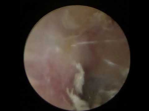
28 year old male patient with an 8 month old hearing loss following blunt trauma over the right ear; an otoscopy was performed showing 30%...
-
Just wanted to share right middle meatus anatomy noticed during nasal endoscopy. 39 year old male patient with 10 year old history of rig...
-
34 year old female patient with a 2 week history of left nasal congestion and obstruction managed with topical steroids. A rigid nasal en...
-
28 year old male patient with an 8 month old hearing loss following blunt trauma over the right ear; an otoscopy was performed showing 30%...


